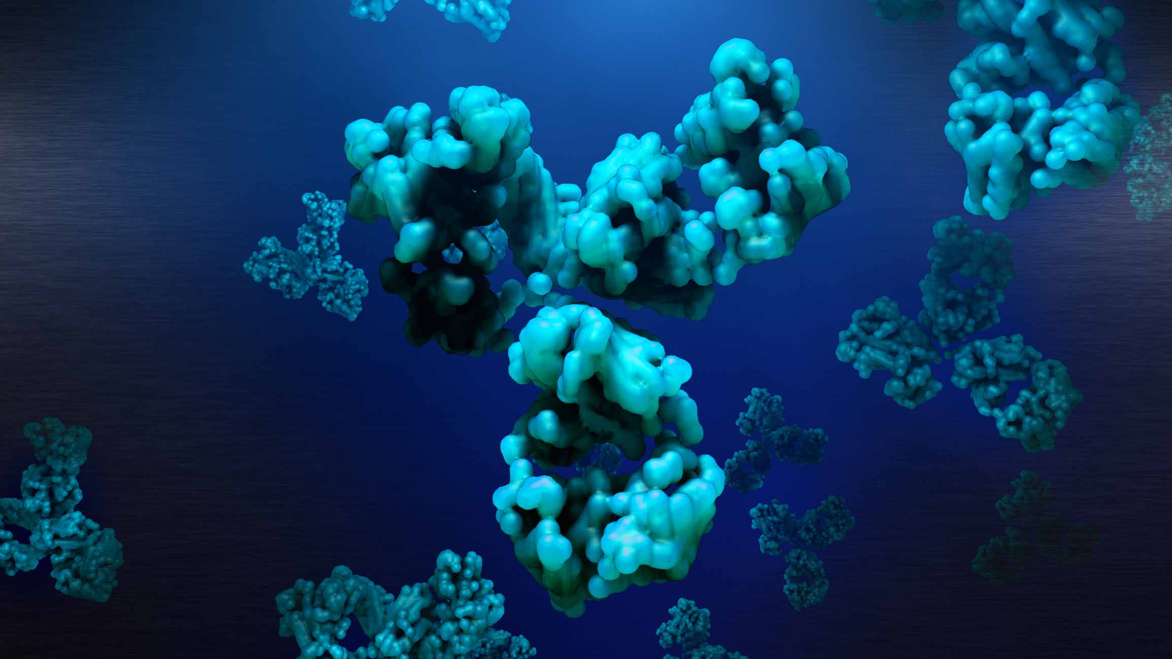
Cryo-EM, or Cryogenic Electron Microscopy, is a cutting-edge imaging technique that enables scientists to visualize molecular structures at near-atomic levels. It’s currently revolutionizing the fields of structural biology, nanotechnology, drug discovery, and beyond.
This article will cover the rise of cryo-EM, the steps in the pipeline, its impacts on various research areas, and the data storage and management challenges associated with it.
The rise of cryo-EM
Over the past two decades, the total number of solved structures added to the Protein Data Bank that were generated by cryo-EM has grown fivefold. The majority of growth, however, has happened in only the past few years.
For quite some time, when it came to determining molecular structures, X-ray crystallography was the main workhorse. This was partially because cryo-EM initially had relatively low resolution capabilities, making it challenging to compete with the high-resolution results obtained from X-ray crystallography.
However, significant improvements in microscopes, detectors, and image processing algorithms sparked rapid improvements in the technique. With an ideal sample, cryo-EM can solve molecular structures with a resolution below 1.5 Å – a level that would have been unthinkable a few years ago.
Cryo-EM now rivals, and in some cases even surpasses, X-ray crystallography in terms of resolution, making it an attractive option for studying a wider range of samples – especially large, dynamic, and non-crystallizable molecules.
The steps in a cryo-EM Pipeline
- Sample preparation: Scientists isolate and purify the molecules of interest, such as proteins or complexes, and carefully preserve them in a near-native state. This is done with a flash freezing process that traps the molecules in a thin layer of vitreous ice at extremely low temps, around -196°C (-321°F).
- Data collection: That frozen sample is loaded into the cryo-electron microscope. Beams of electrons are then fired at it, and the detector beneath captures 2D images of the molecules frozen in various orientations.
- Image processing: The micrographs obtained from step 2 contain useful information about the sample, but they are also noisy and contain artifacts, which brings us to the next step.
- Particle picking: Here, individual molecules within the micrographs need to be identified. Specialized algorithms are used to extract particles from the micrographs. This generates the set of particle images that will be used for 3D reconstruction.
- 3D reconstruction: The set of particle images is then used to calculate a 3D model of the molecule’s structure. Advanced software analyzes and processes the micrographs before combining all the different orientations into a single 3D rendering.
There are a few more steps before the pipeline is complete, including refinement and validation processes. But the end result is a high-resolution 3D model displaying the molecule’s complete structure. Seeing these shapes is vital to understanding how molecules behave, which in turn unlocks the door to new interventions in drug discovery, semi-conductors, energy research, and more.
The impact of cryo-EM
Much of the talk surrounding cryo-EM centers on its applications in drug discovery, and for good reason. By providing detailed insights into the structure of drug targets – particularly membrane proteins, common ones that are notoriously difficult to study using X-ray crystallography – cryo-EM is fast-tracking the development of more effective pharmaceuticals.
But there’s much more to the story. Here are some other areas where cryo-EM promises to make big impacts:
- Energy Research: By providing insights into the structure and function of biomolecules involved in energy conversion processes, cryo-EM can aid in the development of sustainable energy solutions and biofuels. Cryo-EM may also soon help us solve the renewable energy battery storage problem.
- Semiconductors: Cryo-EM is also being utilized in the semiconductor industry to study materials at the atomic level, leading to advancements in nanotechnology and microelectronics. It can provide detailed images of defects and interfaces at the nanometer scale, which is crucial for understanding the properties and performance of semiconductor devices.
- Environmental Science: Cryo-EM aids environmental science research by providing insights into the structure of biomolecules involved in environmental processes. It helps study enzymes involved in bioremediation, nutrient cycling, and pollutant degradation, offering potential sustainability solutions for pressing environmental challenges.
- Neuroscience: Cryo-EM has made significant contributions to neuroscience by helping researchers visualize the complex structures of neurons, synapses, and ion channels. This knowledge is vital for understanding brain function and neurological disorders as well as developing targeted treatments.
As cryo-EM continues to evolve and become more accessible, its impact on various scientific and industrial disciplines will only keep growing.
Cryo-EM data challenges
While Cryo-EM offers exciting possibilities, it also presents significant data storage and management challenges:
- Growing data volumes: Datasets resulting from a single cryo-EM experiment can range from 5 to 40 terabytes, and one microscope can easily generate multiple petabytes of data per year. Scalable storage solutions are essential to accommodate the increasing demands for data storage capacity.
- High data throughput: Cryo-EM instruments capture images at a high rate, producing a continuous stream of data. Data storage solutions need to support high data throughput to avoid bottlenecks during image acquisition, analysis, and storage.
- Long-term data preservation: Cryo-EM data is critical for ongoing research and future reference. To maintain data integrity and accessibility, institutions need reliable, long-term data preservation strategies that ensure data retention for extended periods of time.
- Data redundancy and replication: To prevent data loss due to hardware failures or disasters, data redundancy and replication mechanisms are essential. Cryo-EM facilities should choose storage solutions that integrate fault tolerance and make creating backups and storing copies of data easy.
- Data sharing and collaboration: Cryo-EM is a collaborative field, and researchers often need to share data across different locations. Data storage solutions should facilitate easy and secure data movement.
- Data insights and management: With comprehensive visualization reports clearly showing what data is on their storage, researchers can better understand and manage their data usage and placement.
The bottom line: Cryo-EM pipelines require a scalable, reliable, and high-performance data storage platform capable of handling vastly growing quantities of data.
VDURA Solutions for Cryo-EM
VDURA solutions are deployed at cryo-EM facilities across the globe. Here’s why leading research centers trust VDURA with their workloads:
- Scale limitlessly
And that includes data capacity, performance, and protection. Start with a small entry-point and expand as needed without creating data silos or interrupting workflows.
- Accelerate analysis
High-speed data access lets you quickly retrieve and analyze large datasets and allows multiple teams to work together on the same dataset simultaneously.
- Unlock the full potential of your data
Powerful search and scan capabilities enable you to quickly locate specific datasets and experiments, and an easy-to-use data movement tool lets you manage backup and archive options.
- Experience unparalleled reliability and simplicity
There’s a misconception in the high-performance storage space that you have to compromise system reliability and ease of management to meet your performance needs. VDURA is proof otherwise.
To learn more about how VDURA can optimize your cryo-EM workflows, head over to our life sciences solution page.


